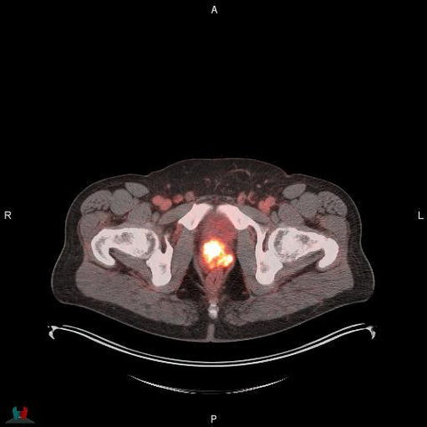
Additional Results of POSLUMA® (Flotufolastat F 18) Performance in Newly Diagnosed, High-risk Prostate Cancer Patients Presented at ASTRO
Additional Results of POSLUMA® (Flotufolastat F 18) Performance in Newly Diagnosed, High-risk Prostate Cancer Patients Presented at ASTRO
− Sub-group analysis from Blue Earth Diagnostics’ Phase 3 LIGHTHOUSE trial highlights POSLUMA utility to guide treatment selection and potentially avoid futile surgery –
This press release is for U.S. audiences only
MONROE TOWNSHIP, N.J. & OXFORD, England--(BUSINESS WIRE)--Blue Earth Diagnostics, a Bracco company and recognized leader in the development and commercialization of innovative positron emission tomography (PET) radiopharmaceuticals, today announced results from a sub-group analysis from the Phase 3 LIGHTHOUSE trial (NC04186819) which evaluated the safety and diagnostic performance of POSLUMA® (flotufolastat F 18) PET in men with newly diagnosed prostate cancer. Specifically, this sub-group examined the performance of flotufolastat F 18 PET in newly diagnosed, high-risk prostate cancer patients who had negative results with conventional imaging. Recently approved by the U.S. FDA, POSLUMA is indicated for positron emission tomography (PET) of prostate-specific membrane antigen (PSMA) positive lesions in men with prostate cancer with suspected metastasis who are candidates for initial definitive therapy or with suspected recurrence based on elevated serum prostate-specific antigen (PSA) level.
Results highlights:
- The sensitivity and specificity of flotufolastat F 18 for the detection of pelvic lymph nodes in 174 patients who underwent surgery ranged from 24-33% and 92-96%, respectively, across 3 blinded, independent readers.
- The detection rate for M1 lesions (lesions that have spread beyond the pelvic area) for flotufolastat F 18 in 197 patients was 14% - 25% across the 3 readers.
- Of identified lesions, 16-25 were successfully verified as True Positive, providing a M1 Verified Detection Rate (VDR) of 8.1 – 13%, across the 3 readers.
“Effective initial staging of prostate cancer, particularly with regards to the detection of metastatic disease, is critical to optimal clinical management of patients,” said Phillip H. Kuo, MD, Ph.D., Departments of Medical Imaging, Medicine, and Biomedical Engineering. “The demonstrated performance of PSMA-PET imaging fits an important unmet need, given that conventional imaging techniques are limited in the information they provide. This analysis from the LIGHTHOUSE study showed that flotufolastat F 18 provides clinically useful information about the presence of both metastatic pelvic lymph nodes and M1 disease prior to surgery in patients with high-risk prostate cancer who had negative results on conventional imaging. Such information can help guide treatment selection and potentially avoid futile surgery for patients with high-risk disease.”
“We are pleased to present these results from the LIGHTHOUSE study to the radiation oncology community at ASTRO,” said David E. Gauden, D.Phil., Chief Executive Officer of Blue Earth Diagnostics. “POSLUMA provides physicians with high-quality diagnostic information based on its diagnostic performance even at low PSA levels, high-affinity PSMA binding and low urinary bladder activity. In a post-hoc Phase 3 analysis, as well as in preclinical and Phase 1 studies, POSLUMA demonstrated low urinary bladder activity, providing for enhanced image evaluation in the prostate and regions near the ureters for patients with prostate cancer. The product is labeled with the radioisotope fluorine-18 (18F) to leverage the high image quality of 18F-labeled PSMA PET imaging, to facilitate effective detection of disease and enable broad, readily available geographic access for patients through the manufacturing and distribution network of our commercial U.S. manufacturer and distributor, PETNET Solutions Inc, A Siemens Healthineers Company. Nationally recognized clinical oncology guidelines for prostate cancer now include POSLUMA, alongside and for all the same categories as the other currently FDA-approved PSMA PET radiopharmaceuticals.”
The findings were discussed in a poster Q&A session at the 2023 ASTRO Annual Meeting on October 3, 2023,“Diagnostic Performance of 18F-rhPSMA-7.3 PET in Men with Newly Diagnosed High-risk Prostate Cancer and Negative Conventional Imaging,” by Phillip H. Kuo, MD, Ph.D., Departments of Medical Imaging, Medicine, and Biomedical Engineering, University of Arizona, Tucson, Ariz. on behalf of Gary A. Ulaner, MD, Ph.D., Hoag Family Cancer Institute, Irvine, Calif. and University of Southern California, Los Angeles, Calif., for the LIGHTHOUSE Study Group. Full session details and the abstract are available in the ASTRO online program here.
About the study
The sub-group analysis of LIGHTHOUSE data evaluated a subgroup of patients with high/very high-risk prostate cancer who had no evidence of nodal or metastatic disease on conventional imaging. Treatment-naïve patients scheduled for radical prostatectomy (RP) plus pelvic lymph node (PLN) dissection underwent flotufolastat F 18 PET. Local readers interpreted the scans prior to RP and ahead of a blinded read by 3 central readers. If the local read indicated M1 disease, verification (biopsy, surgery, or confirmatory follow-up imaging) of PET-positive M1 lesions was attempted before treatment. The analysis evaluated flotufolastat F 18 sensitivity and specificity for detection of PLN metastases in all high/very high-risk patients with negative conventional imaging at baseline who underwent flotufolastat F 18 PET and subsequent surgery. Histopathology was used as the standard of truth (SoT). Additionally, the M1 verified detection rate (VDR; % of patients with true positive (TP) M1 lesions using histopathology or follow-up imaging as SoT out of all patients scanned) was assessed in an extended population of all patients who had flotufolastat F 18 PET, regardless of surgery.
The sensitivity and specificity for PLN detection among 174 men with very/high-risk PCa and negative conventional imaging ranged from 24-33% and 92-96%, respectively, across the 3 readers. M1 lesions were identified in 28-51 of the 197 patients in the extended population, giving an overall M1 detection rate of 14-26%, across the 3 readers. Of the identified lesions, 16-25 were successfully verified (predominantly using follow-up imaging as SoT) as TP, providing a M1 VDR of 8.1-13%, across the 3 readers.
No serious adverse events were observed in the LIGHTHOUSE study. Overall, 28 of the 356 (7.9%) patients who received flotufolastat F 18 had at least one treatment-emergent adverse event that was considered possibly related to flotufolastat F 18. The most frequently reported adverse event for patients in the LIGHTHOUSE study was injection site pain among 0.8% (3/356) of patients.
About Radiohybrid Prostate-Specific Membrane Antigen (rhPSMA)
Radiohybrid Prostate-Specific Membrane Antigen (rhPSMA) compounds consist of a radiohybrid (“rh”) Prostate-Specific Membrane Antigen-targeted receptor ligand which attaches to and is internalized by prostate cancer cells, and they may be radiolabeled with imaging isotopes for PET imaging, or with therapeutic isotopes for therapeutic use – providing the potential for creating a true theranostic technology. Radiohybrid technology and rhPSMA originated from the Technical University of Munich, Germany. Blue Earth Diagnostics acquired exclusive, worldwide rights to rhPSMA diagnostic imaging technology from Scintomics GmbH in 2018, and therapeutic rights in 2020, and sublicensed the therapeutic application to its sister company Blue Earth Therapeutics. Blue Earth Diagnostics received U.S. Food and Drug Administration approval for its radiohybrid PET diagnostic imaging product for use in prostate cancer in 2023. rhPSMA compounds for potential therapeutic use are investigational and have not received regulatory approval.
Indication and Important Safety Information About POSLUMA
INDICATION
POSLUMA® (flotufolastat F 18) injection is indicated for positron emission tomography (PET) of prostate-specific membrane antigen (PSMA) positive lesions in men with prostate cancer
- with suspected metastasis who are candidates for initial definitive therapy
- with suspected recurrence based on elevated serum prostate-specific antigen (PSA) level
IMPORTANT SAFETY INFORMATION
- Image interpretation errors can occur with POSLUMA PET. A negative image does not rule out the presence of prostate cancer and a positive image does not confirm the presence of prostate cancer. The performance of POSLUMA for imaging metastatic pelvic lymph nodes in patients prior to initial definitive therapy seems to be affected by serum PSA levels and risk grouping. The performance of POSLUMA for imaging patients with biochemical evidence of recurrence of prostate cancer seems to be affected by serum PSA levels. Flotufolastat F 18 uptake is not specific for prostate cancer and may occur in other types of cancer, in non-malignant processes, and in normal tissues. Clinical correlation, which may include histopathological evaluation, is recommended.
- Risk of Image Misinterpretation in Patients with Suspected Prostate Cancer Recurrence: The interpretation of POSLUMA PET may differ depending on imaging readers, particularly in the prostate/prostate bed region. Because of the associated risk of false positive interpretation, consider multidisciplinary consultation and histopathological confirmation when clinical decision-making hinges on flotufolastat F 18 uptake only in the prostate/prostate bed region or only on uptake interpreted as borderline.
- POSLUMA use contributes to a patient’s overall long-term cumulative radiation exposure. Long-term cumulative radiation exposure is associated with an increased risk for cancer. Advise patients to hydrate before and after administration and to void frequently after administration. Ensure safe handling to minimize radiation exposure to the patient and health care providers.
- The adverse reactions reported in ≥0.4% of patients in clinical studies were diarrhea, blood pressure increase and injection site pain.
- Drug Interactions: androgen deprivation therapy (ADT) and other therapies targeting the androgen pathway, such as androgen receptor antagonists, may result in changes in uptake of flotufolastat F 18 in prostate cancer. The effect of these therapies on performance of POSLUMA PET has not been established.
To report suspected adverse reactions to POSLUMA, call 1-844-POSLUMA (1-844-767-5862) or contact FDA at 1-800-FDA-1088 or www.fda.gov/medwatch.
Full POSLUMA prescribing information is available at www.posluma.com/prescribing-information.pdf.
About Blue Earth Diagnostics
Blue Earth Diagnostics, an indirect subsidiary of Bracco Imaging S.p.A., is a growing international molecular imaging company focused on delivering innovative, well-differentiated diagnostic solutions that inform patient care. Formed in 2014, the Company’s success is driven by its management expertise and supported by a demonstrated track record of rapid development and commercialization of positron emission tomography (PET) radiopharmaceuticals. Blue Earth Diagnostics’ expanding oncology portfolio encompasses a variety of disease states, including prostate cancer and neuro-oncology. Blue Earth Diagnostics is committed to the timely development and commercialization of precision radiopharmaceuticals for potential use in imaging and therapy. For more information, please visit: www.blueearthdiagnostics.com.
About Bracco Imaging
Bracco Imaging S.p.A., part of the Bracco Group, is a world-leading diagnostic imaging provider. Headquartered in Milan, Italy, Bracco Imaging develops, manufactures and markets diagnostic imaging agents and solutions. It offers a product and solution portfolio for all key diagnostic imaging modalities: X-ray imaging (including Computed Tomography-CT, Interventional Radiology, and Cardiac Catheterization), Magnetic Resonance Imaging (MRI), Contrast Enhanced Ultrasound (CEUS), and Nuclear Medicine through radioactive tracers and novel PET imaging agents to inform clinical management and guide care for cancer patients in areas of unmet medical need. Our continually evolving portfolio is completed by a range of medical devices, advanced administration systems and dose-management software. In 2019 Bracco Imaging enriched its product portfolio by expanding the range of oncology nuclear imaging solutions in the urology segment and other specialties with the acquisition of Blue Earth Diagnostics. In 2021, Bracco Imaging established Blue Earth Therapeutics as a separate, cutting-edge biotechnology vehicle to develop radiopharmaceutical therapies. Visit: www.braccoimaging.com.
Contacts
For Blue Earth Diagnostics (U.S.)
Priscilla Harlan
Vice President, Corporate Communications
(M) (781) 799-7917
priscilla.harlan@blueearthdx.com
For Blue Earth Diagnostics (UK)
Clare Gidley
Associate Director Marketing and Communications
Tel: +44 (0) 7917 536939
clare.gidley@blueearthdx.com
Media
Sam Brown Inc.
Mike Beyer
(M) (312) 961-2502
mikebeyer@sambrown.com


