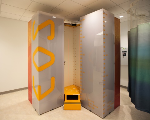
EMERYVILLE, Calif.--(BUSINESS WIRE)--A low-dose radiation X-ray technology that takes high-quality, full-length images of a child in a standing position is now available at Stanford Children’s Health Specialty Services Emeryville.
The orthopedic imaging scanner, manufactured by EOS imaging, is the first and only of its kind in Northern California. It represents another step in the mission of Stanford Children’s Health to make high-quality care more accessible in outpatient settings and near where families live. This expanded access reduces costs, increases convenience and creates a better patient experience.
That experience includes safer imaging, said orthopedic surgeon James Policy, MD, noting that the typical radiation dose from the new technology is between one-fifth and one-third of the exposure from a standard digital X-ray.
“This is the kind of imaging you want for a child that has scoliosis and will need X-rays multiple times a year for many years,” said Policy, who is also a clinical assistant professor at the Stanford University School of Medicine.
That’s the situation faced by 13-year-old scoliosis patient Chaeli Borchers of Walnut Creek, who has received many conventional X-rays in her life. “When I heard this new low-dose alternative was available,” said mom Gina, “we jumped at it.” Chaeli had her first session in December. “It was great,” added mom, “and I won’t take her anywhere else in the future.”
Another advantage is achieving seamless, full-length images of a patient in a standing position. “For scoliosis and for leg-length X-rays, or to look for angular deformities in the legs, you definitely want the child standing,” Policy said.
The whole process takes only four to six seconds, Policy said. The Emeryville team has successfully used the technology with children as young as 18 months. The speed of the imaging helps keep radiation exposure low, not to mention minimizing the strain of making an active child stand still.
“For patients requiring spine X-rays and extremity images, we’re now capable of outstanding imaging at a substantially reduced radiation dose,” said Richard Barth, MD, chief of pediatric radiology at Lucile Packard Children’s Hospital Stanford and professor of radiology at the School of Medicine.
Policy emphasized that the new technology is not replacing conventional digital X-rays, which are used when a standing image isn’t needed, such as for fractured wrists or most other broken bones.
Ensuring that this type of imaging is available in a central location like Emeryville provides convenience, safety and world-class, nurturing care for patients throughout the region. It’s another important way Stanford Children’s Health is expanding access to its network of care for children and expectant mothers.


