LOS ANGELES--(BUSINESS WIRE)--A mother and her 25-week-old fetus are doing well after a team of physicians performed a successful in utero cardiac interventional procedure on the fetus at CHA Hollywood Presbyterian Medical Center late last month.
The minimally invasive procedure, known as a fetal aortic valvuloplasty, was a first for a Southern California hospital. Designed to treat a congenital heart defect known as critical aortic stenosis and evolving hypoplastic left heart syndrome, doctors succeeded in using a tiny balloon to open the fetus’s narrow aortic valve in order to increase blood flow to the body, improve left heart function and promote normal left heart growth during the critical third trimester growing stage.
“Right now mom and baby are doing well,” says surgeon Ramen H. Chmait, M.D., director of Los Angeles Fetal Therapy at CHA Hollywood Presbyterian Medical Center, Children’s Hospital Los Angeles and the USC Institute for Maternal-Fetal Health since 2006. “We are optimistic that the baby’s heart will be much better off because of this procedure."
Chmait performed the procedure on Sept. 25 with Children’s Hospital Los Angeles pediatric interventional cardiologist Frank F. Ing, M.D., FAAP, FACC, FSCAI. Collaborating for the first time, both physicians had performed the delicate fetal procedure before at hospitals outside of California. “The surgery went beautifully; we were able to open up the aortic valve,” says Ing, associate chief of the Division of Cardiology and director of the Cardiac Catheterization Laboratory at Children’s Hospital. “Fetal cardiac intervention is a relatively new field and requires expertise, commitment and collaboration among four specialty areas: pediatric interventional cardiology, fetal echocardiography, maternal-fetal-medicine and anesthesia.”
The doctors emphasized that the partnership between the specialists from Children's Hospital, the University of Southern California and CHA Hollywood Presbyterian was critical to the procedure’s success.
Left untreated, critical aortic stenosis results in a severely damaged left ventricle in newborn infants and can sometimes progress to a dangerous condition called hypoplastic left heart syndrome (HLHS) when the child is born. HLHS can be fatal and typically requires three separate and very risky surgeries to correct after birth.
"In fetuses with critical aortic stenosis, the aortic valve is very narrow,” says Chmait, who explained the heart defect in the fetus was originally diagnosed by an echocardiogram earlier in the pregnancy. The condition “prevents normal emptying of blood from the left ventricle to the aorta. If treatment is delayed until after birth, the left ventricle can become so damaged that it cannot function normally.”
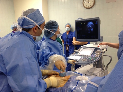
The patient, a 28-year-old Sylmar, Calif. mother of two, was 25 weeks pregnant at the time of procedure, which took a total of three hours and involved a team of 12, including physicians and support staff. Much of that time was devoted to Chmait’s maneuvering the fetus into position by manually adjusting the mother’s abdomen. With the assistance of a detailed ultrasound imaging by Children’s Hospital fetal cardiologist Jay Pruetz, M.D., Chmait inserted a special needle into the womb, through the fetal chest and into the peanut-sized heart’s left ventricle, positioning it below the aortic valve. As Chmait steadied the needle, Ing used Chmait’s hand as a platform for his hand to thread a hair-thin wire through the needle and out the tip, positioning the wire across the fetus’s aortic valve. The wire was then used as a rail for Ing to maneuver a tiny balloon-tipped catheter into position across the valve. The balloon was carefully inflated by Ing, opening the valve and increasing blood flow into the aorta. The balloon, wire and needle were then removed.
The time from actual needle insertion to the balloon opening took roughly 15 minutes, Ing says. The mother was sedated during the procedure.
“After the procedure, blood was able to move easily across the valve and into the aorta,” Chmait says.
The fetal aortic valvuloplasty procedure, first performed in England in 1991, has been completed more than 100 times total in the U.S., with most of those surgeries taking place at Boston Children’s Hospital. Only a handful of hospitals west of the Mississippi River have performed the procedure.
Not only does the rare procedure give the child better heart function in utero, but it also allows for improved left heart growth and thus increases the chances of the baby having a normal two-ventricle system, which would not happen if left untreated, Pruetz explained. Even if the left ventricle doesn’t grow into an equal partner with the right ventricle, studies show heart performance is improved. “Given the choice between having two engines, one of which is totally too small versus having two engines and one is smaller but useable, I’d rather have the latter,” Ing says.
About CHA Hollywood Presbyterian Medical Center
CHA Hollywood Presbyterian Medical Center is an acute care facility that has been caring for the Hollywood community and surrounding areas since 1923. The hospital is committed to serving local multicultural communities with quality medical and nursing care. With more than 500 physicians representing virtually every specialty, CHA Hollywood Presbyterian Medical Center is a 429 bed facility located in Los Angeles, Calif. that provides a full range of acute services including fetal therapy services.
For more information, visit www.hollywoodpresbyterian.com or www.LosAngelesFetalTherapy.org
About Children’s Hospital Los Angeles
Children's Hospital Los Angeles has been named the best children’s hospital on the West Coast and among the top five in the nation for clinical excellence with its selection to the prestigious US News & World Report Honor Roll. Children’s Hospital is home to The Saban Research Institute, one of the largest and most productive pediatric research facilities in the United States. Children’s Hospital is also one of America's premier teaching hospitals through its affiliation since 1932 with the Keck School of Medicine of the University of Southern California.
For more information, visit CHLA.org. Follow us on Twitter, Facebook, YouTube and LinkedIn, or visit our blog: WeTreatKidsBetter.org.
About Keck Medicine of USC
Keck Medicine of USC is the University of Southern California's medical enterprise, one of only two university-owned academic medical centers in the Los Angeles area. Encompassing academic, research and clinical entities, it consists of the Keck School of Medicine of USC, one of the top medical schools in Southern California; the renowned USC Norris Comprehensive Cancer Center, one of the first comprehensive cancer centers established in the United States; the USC Care faculty practice; the Keck Medical Center of USC, which includes two acute care hospitals: 411-bed Keck Hospital of USC and 60-bed USC Norris Cancer Hospital; and USC Verdugo Hills Hospital, a 158-bed community hospital. It also includes outpatient facilities in Beverly Hills, downtown Los Angeles, La Cañada Flintridge, Pasadena, and the USC University Park Campus. USC faculty physicians and Keck School of Medicine departments also have practices throughout Los Angeles, Orange and Riverside counties.


